ECG Lead Placement: 12-Lead Electrode Positions Explained
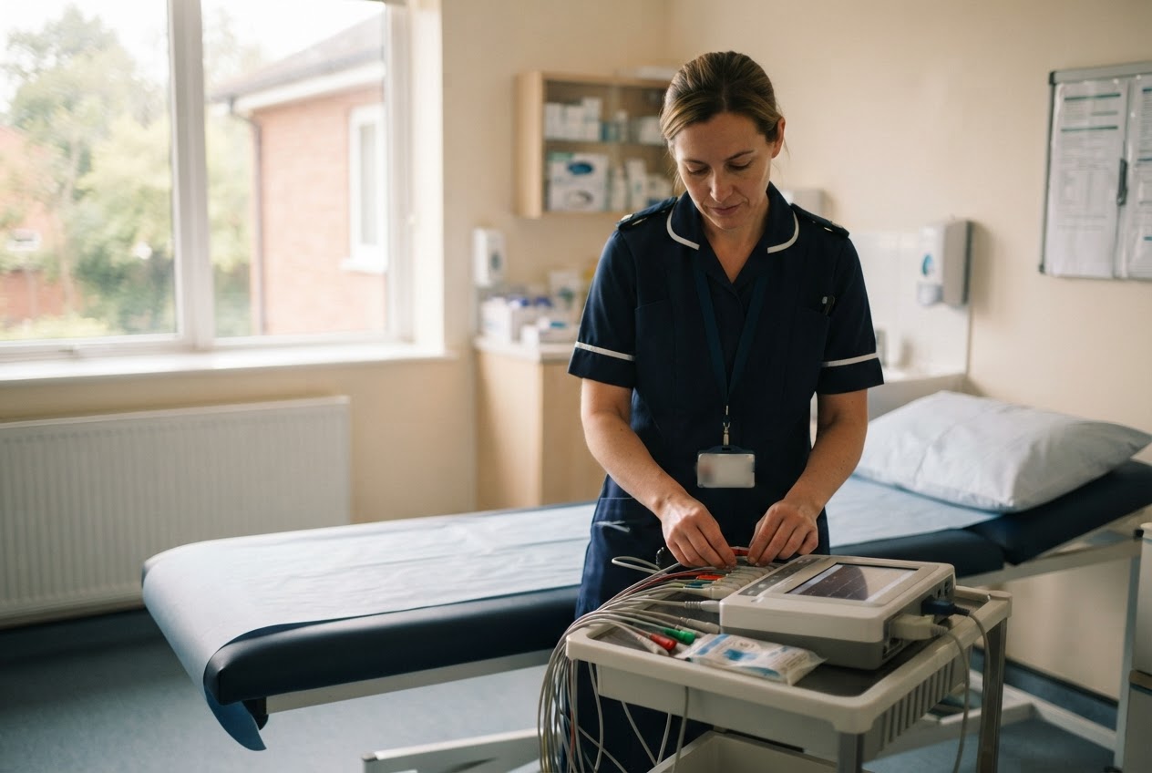

A standard 12-lead ECG uses 10 electrodes: four on the limbs and six on the chest. The limb electrodes go on the forearms just above the wrists and the lower legs just above the ankles. The chest electrodes run from V1, in the fourth intercostal space at the right edge of the breastbone, round to V6 in the left mid-axillary line.
This guide follows the UK standard: the Society for Cardiac Science and Technology (SCST) guideline on recording a standard 12-lead ECG, updated in September 2024. It's a reference for nurses and clinical staff. Always follow your local policy, and practise under supervision before recording ECGs on your own.
Where the 10 electrodes go
The "12 leads" are views of the heart's electrical activity that the machine builds from these 10 electrodes. Getting each electrode in the right place is what makes those views reliable.
Limb electrodes
- RA (usually red): right forearm, just above the wrist
- LA (usually yellow): left forearm, just above the wrist
- LL (usually green): left lower leg, just above the ankle
- RL (usually black): right lower leg, just above the ankle
Colours follow international recommendations but can vary by manufacturer, so check the labels on your machine. The SCST says limb electrodes should only move further up the limbs for a significant tremor or an amputation, and any non-standard position must be written on the recording. They must not go on the torso, because that significantly changes the size of the waves.
Chest electrodes (V1 to V6)
- V1: fourth intercostal space, right edge of the sternum
- V2: fourth intercostal space, left edge of the sternum
- V3: midway between V2 and V4
- V4: fifth intercostal space, in line with the middle of the left collarbone (mid-clavicular line)
- V5: left anterior axillary line, level with V4
- V6: left mid-axillary line, level with V4 and V5
Finding the right space
The SCST method starts from the angle of Louis, the bony ridge where the top part of the breastbone meets the main part. Beside it is the second intercostal space. From there you count down the spaces to the fourth for V1, then count again on the left for V2. The guideline warns that rib spaces aren't always level on both sides, so count on each side rather than placing V2 opposite V1 by eye. Place V4 next, then V3 between V2 and V4, then V5 and V6 level with V4.
Common placement errors and their effect
The SCST says V1 and V2 are frequently placed too high, and V4, V5 and V6 too low. Both errors can cause diagnostically misleading changes to the ECG. Other problems that show up on the trace:
- Limb electrodes on the chest or shoulders: changes the size of the waves.
- Arm leads swapped: changes how the limb leads look and can mislead whoever reviews the trace. Check each lead against its label before you record.
- Chest electrodes placed by eye: the SCST method relies on counting the spaces, and guessing makes misplacement more likely.
- A tense or shivering person: muscle activity causes interference that makes the ECG harder to read.
- Unlabelled changes: a non-standard position the reviewer doesn't know about can be mistaken for a real change.
Preparing the person
A good trace starts with a relaxed, comfortable person who knows what's happening.
- Explain and get consent. The SCST says verbal consent is acceptable for an ECG. In England and Wales, if someone can't consent, follow the Mental Capacity Act 2005 and your local policy. See our Mental Capacity Act training.
- Protect their dignity. Clothing above the waist usually needs to come off for the chest electrodes. Give privacy, and cover the person once the cables are on.
- Offer a chaperone. The SCST recommends a formal clinical chaperone and says a relative or carer isn't ideal in that role. If the person declines, record the offer and their answer.
- Position them well. Lying back at about 45 degrees is recommended, with arms and legs supported. If they're recorded in another position, such as in a wheelchair, note it on the ECG.
- Prepare the skin. Clean with mild soap and water or an alcohol wipe; very light abrasion may be needed. If hair must be removed, get verbal consent first and use a single-use blade. Take care with fragile skin.
Breast tissue
Where breast tissue covers the correct positions, the convention is to place V4 to V6 beneath the left breast. The SCST suggests lifting the breast with care using the back of the hand, to keep contact to a minimum. Count the spaces as usual rather than placing electrodes on top of breast tissue.
Recording a good trace
- Ask the person to lie still, relax and not talk while the machine records.
- Check every electrode is stuck down and no cable is pulling on it.
- Keep the machine on the standard settings your local policy sets. If you have to change the gain or use a filter to reduce interference, the SCST says this must be marked on the ECG.
- Label the recording with at least two identifiers, such as name and date of birth, plus the date and time and who recorded it.
- Note any symptoms at the time, such as chest pain, and any non-standard electrode positions.
- Pass the ECG to the appropriate clinician promptly. Recording a trace isn't the same as interpreting it.
If the person is unwell, for example with chest pain, breathlessness or collapse, don't let the ECG delay emergency help. Follow your emergency procedure. Our Basic Life Support training covers what to do if someone collapses, and Immediate Life Support is there for clinical teams who need more.
Who should record ECGs
ECGs are usually recorded by nurses and other suitably qualified staff, after training and a sign-off in practice under your local policy. In England, CQC's Regulation 12 expects the people giving care and treatment to have the qualifications, skills and experience to do so safely. A poorly recorded ECG can mislead the clinician reviewing it, so the recording itself matters. Care regulators in Scotland, Wales and Northern Ireland expect the same.
How Kasorb can help
Kasorb's ECG training teaches nurses and clinical staff to record a 12-lead ECG correctly: lead locations and placement, the recording process and recognising a good trace, plus the basics of reviewing results and common arrhythmias. Staff practise placing leads and recording ECGs on a manikin with a 12-lead ECG machine, with feedback from the trainer.
It runs on-site anywhere in the UK, as a 3-hour awareness session or a full day, for up to 12 staff at one price for the group. Your trainer has used these skills for real. It's awareness-level training, not diagnostic interpretation, which stays with the appropriate clinician. Certificates are emailed the next day.
FAQs
Where do the leads go on a 12-lead ECG?
Four limb electrodes go on the forearms just above the wrists and the lower legs just above the ankles. Six chest electrodes go from V1 and V2 in the fourth intercostal space either side of the sternum, through V3 and V4, to V5 and V6 level with V4 along the left side of the chest.
Where does V1 go on an ECG?
V1 goes in the fourth intercostal space at the right edge of the sternum. Find the angle of Louis, the ridge near the top of the breastbone, move to the second space beside it and count down to the fourth. The SCST says V1 is often placed too high, so count rather than guess.
What happens if ECG leads are placed wrong?
Misplaced electrodes change the shape and size of the waves, which can mislead the clinician reviewing the ECG. The SCST says chest electrodes placed too high or too low can cause diagnostically misleading changes. Limb electrodes on the torso change wave size, and swapped arm leads alter the limb leads.
Where do you put ECG leads on a woman?
The positions are the same. Where breast tissue covers the correct spots, the SCST convention is to place V4, V5 and V6 beneath the left breast, lifting it gently with the back of the hand. Offer a chaperone, give privacy and cover the person once the cables are attached.
Can you put ECG limb leads on the shoulders?
Not for a standard 12-lead ECG. The SCST says limb electrodes must not be placed on the torso, because this significantly alters wave size. They go just above the wrists and ankles, and only move further up the limbs for a significant tremor or amputation, with the change written on the recording.
On-site training for your whole team at one flat rate. See what's covered, prices and how to book.
See the course

Not sure which course your team needs? Tell us about your setting and we'll confirm within 48 hours.